
A Closer Look Inside a Sheep Brain
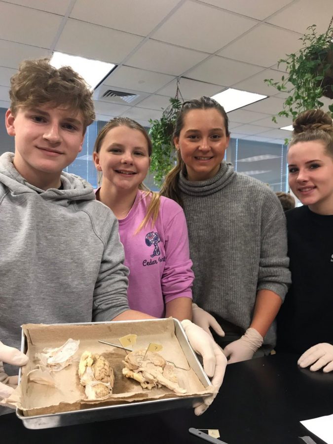
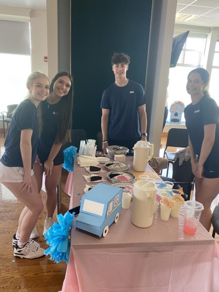
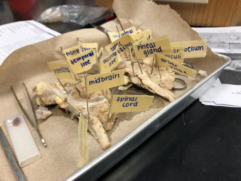
Ethan Apple, Jillian Bowman, Molly Bobik, and Kylie Blaszkowski showcase the sheep brain they dissected in anatomy class.
What would we do without our brains? How would we process anything? This week, students in the anatomy classes dissected a sheep brain. A once in a lifetime experience presented itself recently to many young students who may be doing something along the lines of this activity in their future endeavors.
Students in small groups dissected a sheep brain and got up close and personal with its structure and unique design. With each type of brain, there are differences in size, structure, and brain regions, but the general structures and the area of location are relatively the same or very similar to the human brain.
The sheep brains come from a biological supply company and this year the school received 28 of them for the classes. During this lab, a sheep brain was cut open and all the divided structures that allow a sheep to survive and sustain life when everything worked as one were labeled. By participating in this hands on experiment, students are able to understand how the brain works and what makes the human brain stand out from other organisms. “I thought it was interesting learning how all the parts work together,” said senior Eve O’Sullivan.
“I have done the lab for the last few years, especially as I have tried to make the brain unit more intensive,” said Mrs. LeVan.
The sheep brain dissection allowed familiarization with the three dimensional structure of the brain. One of the most important concepts of this lab is realizing how the anatomy precedes the physiology. The structure of how the brain is put together explains how it works and what this means about the operation of the brain. The observation and evolution indicate that there are many similarities between the sheep brain and the human brain.
“When you are holding a fixed brain in your hand, the diagrams you study and pictures in the book come to life,” said LeVan.
The minor differences, however, are instructive. The pre-lab work was critical for understanding and being able to locate important structures in the sheep brain. By comprehending the general concept, students related the different structures of the brain, especially once they started cutting it open.
I liked seeing all the structures that you can’t see from the outside.
— Eve O'Sullivan
When it was time to dissect, the students put on gloves and got right into the dissection using scissors, tweezers, and a scalpel. They first began removing the layers of the meninges, which are the protective coverings on the outside of the brain. The dura mater, the tough layer, was almost entirely removed before the actual dissection started.
Once the dura mater was removed, the students cut down the medial longitudinal fissure, which separates the right and left hemispheres of the cerebrum. A total of twenty-two parts of the brain were labeled while the students placed pins into the sheep brain to show where each one was located. Since the brain was on a dissection tray and the groups were small, the different parts were easily able to be seen. “I think it is helpful because I can see what it actually looks like instead of just on a diagram,” said O’Sullivan.
“This lab is beneficial because the students are practicing dissection,” said LeVan. “Dissection is a skill that needs practiced and anytime there is an opportunity to incorporate it in the curriculum, I take advantage.”
The opportunity to dissect a sheep brain taught what makes the human brain stand out in comparison to the sheep brain. Due to the fact our brain is several times larger than a sheep’s brain, this allows us to do many more functions that are not possible for any other organism on this planet. Because of more folds in the human brain, this allows us to carry a lot more information compared to a sheep brain with much less. The human brain gives the ability to talk and to think before we act while most organisms react from instinct.
The students were able to gain knowledge that they can not only use inside the classroom, but beyond too. “I think that this lab will help me in the future because I’ll be a step ahead in college,” said O’Sullivan.

I am currently a Senior at Greater Latrobe Senior High School and hope to pursue a career in the medical field after high school. I am still in the process...